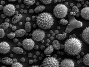
The electron microscope (EM) was first tested by Max Knoll and Ernst Ruska at the Berlin Technische Hochschule in 1931, remarkably overcoming the resolution limits of visible light for the first time. Modern electron microscopes can magnify objects up to 10 million times their size, and have been used extensively to visualize the inner workings of cells. However, biological imaging by electron microscopy (EM) is usually limited to producing grayscale images since its invention.

In the past 15 years, material scientists have developed methods of visualizing colored EM images, but their utility has been limited to imaging synthetic matter. However, a recent study, published in Cell Chemical Biology, has demonstrated a new method to bring super-high resolution colored imaging to the study of biology. The technique uses a special stain made from two rare earth metals, which label the molecules in cells on a microscope slide. When an electron beam is applied to the slide, the majority of electrons pass easily through the sample. However, some electrons that encounter the rare earth metal atoms lose energy, and these low-energy electrons can be detected by a filter on the microscope. Each of the two metals produces a different color. By overlaying the images of the cells before and after labeling, scientists can now create colored images to see details not visible in black and white images.
The study paves the way for scientists to visualize how drugs are delivered to cells, and also how proteins travel within the cell. The developers intend to add additional layers of metals, enabling the colorization of more molecules. By increasing the number of detectable colors, this technique could be used to study even more complex biological processes.
Peer edited by Chiungwei Huang.
Follow us on social media and never miss an article: