 Hayli Spence-Osorio
The blade of the cryostat makes a satisfying zwhoop as it slices through the frozen chunk of mouse brain tissue in front of me. A cryostat is a common enough piece of lab equipment; it maintains sub-zero temperatures while frozen tissue samples, like this mouse brain, are thinly sliced by researchers such as myself. Since the tissue is frozen, it’s extremely solid and durable. This allows me to collect tissue sections thinner than a single human hair every time the blade slices through the tissue. I gently pick up a tissue slice with metal forceps and drop it into a well plate filled with viscous cryoprotectant – a solution that will preserve my samples in a freezer for years to come. Dozens of equally thin tissue sections float in the wells, all containing a fascinating brain structure called the visual cortex.
All brains have a cortex. This is the outer “shell” of the brain, wrapped snugly around midbrain regions responsible for collecting and relaying sensory information to the cortex. The cortex then collects this information, compiles it, and executes cognitive responses. This allows us to perform complex decision making in response to environmental cues. The visual cortex is a specialized cortical region that processes information entering the visual system through your eyes. One of the most interesting and critical functions of the visual cortex is that this specialized cortical region is entirely responsible for flipping visual information on its head and making sure we see the world around us right-side up.
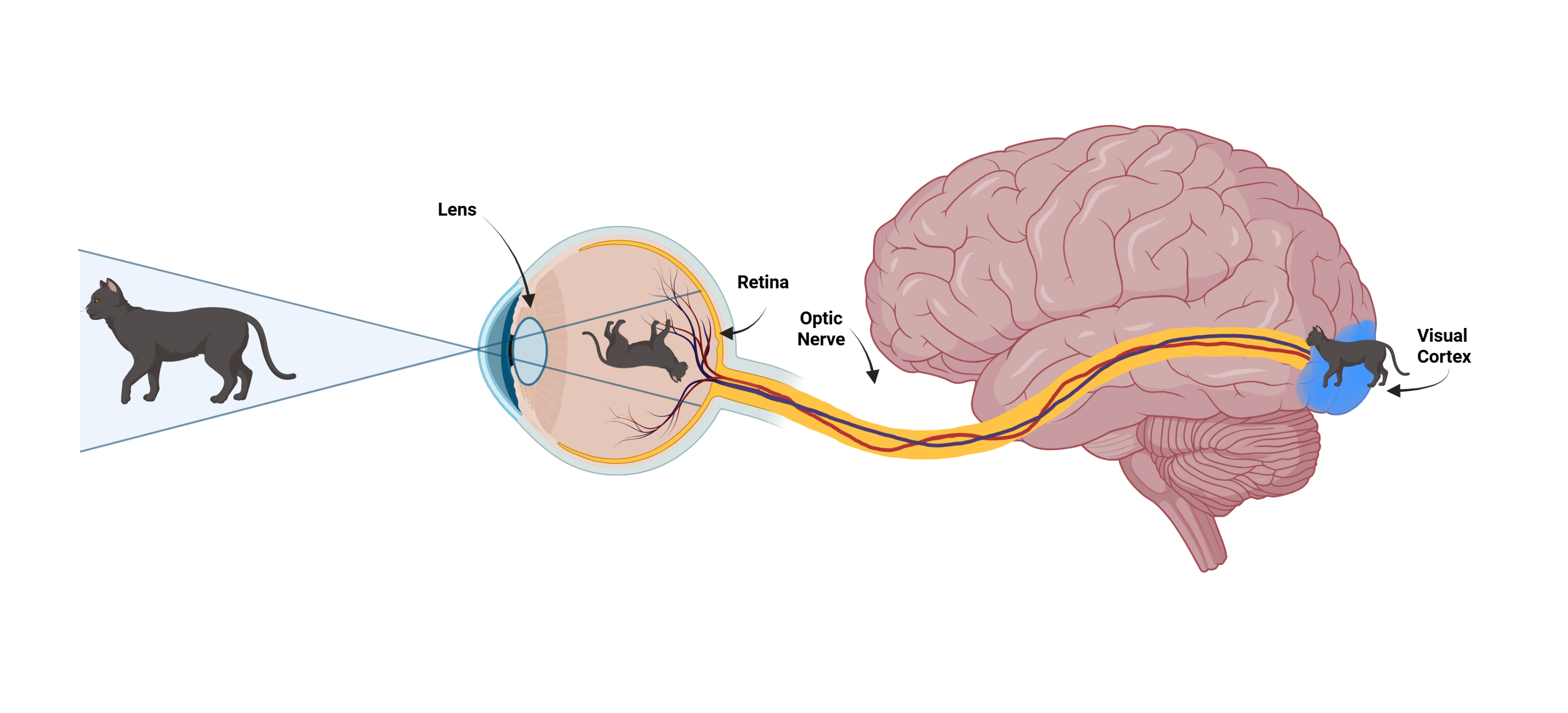
Without the visual cortex, we and our animal friends would actually perceive the world upside down. Eyes act as lenses, collecting and focusing light reflected from the objects around us onto the retina. When light passes through a lens, the image is reflected through a focal point and flipped upside down. This upside-down image is collected by specialized cells in your retina. Interestingly, even the retina is upside-down! There are layers of cells in the retina, and the cells that capture light are in the bottom layer beneath the other cells. Once these cells capture light, they send information about the light through the layers of cells to the outermost layer. This final layer of cells sends long processes (think cables) through the optic nerve. The signal is sent along the optic nerve to the visual cortex. There, in the back of the brain, visual information is processed. So, in some ways, your eyes really are in the back of your head! Once the visual cortex figures out what the “image” of the environment is, it flips it right-side up, and that final, edited image is what you perceive. All of this happens instantaneously, ensuring that you never have to navigate the world upside-down.
You would think the visual system would have evolved to flip the incoming images right-side up a little faster – maybe even in the retina itself. But it didn’t, and the fact that the world we see and perceive through our eyes is something our brains actually create in the backs of our heads is infinitely cool, if a bit confusing at times.
I finish collecting the brain tissue I need and lock the cryostat blade into place. I can’t imagine slicing brain tissue upside down and still keeping all of my fingers intact. Thanks to my visual cortex, I don’t have to!
Peer Edited by: Claire Miller
