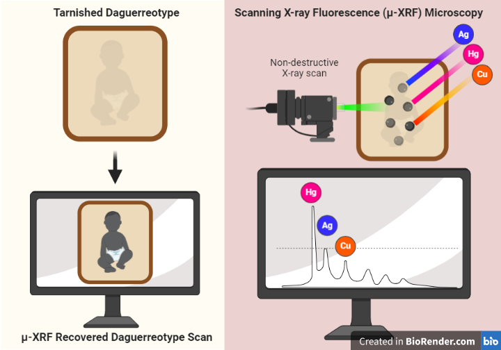
To the rejoice of art conservationists and scientists alike, a team of researchers from the University of Western Ontario, in collaboration with the National Gallery of Canada, the Canadian Light Source facility, and the Cornell High Energy Synchrotron Source, have successfully developed a technique using synchrotron-based scanning X-ray fluorescence (μ-XRF) microscopy to recover heavily degraded daguerreotypes.
Daguerreotypes were the first publicly accessible and commercialized form of photography, though the cost likely limited access to the middle and upper class. Daguerreotypy was invented by the French Louis-Jacques-Mandé Daguerre, after which the technique was named, and Nicéphore Niépce in the 1830s. These photographs were highly popular between 1840-50s prior to the invention of more long-lasting forms of photography using the wet collodion process (tintypes and ambrotypes, anyone?).
What are the principles of this photographic technique?
Daguerreotypes are copper plates coated with a highly polished surface of silver, which was sensitized to light exposure using iodide fumes. Once exposed to light during the photoshoot, the copper plate is quickly developed with mercury vapor and fixed using a common salt solution. The resulting images are very sensitive, any exposure to air will start to tarnish the silver; hence why these photographs are often found in protective cases of leather, covered by glass and lined with silk or velvet (see images below). Exposure times for earlier daguerreotypes varied between 3-20 minutes, a whopping improvement from the earliest photograph recorded, a result of an 8 hour exposure period.
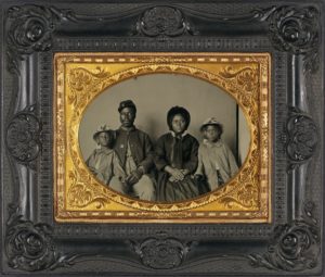
Unidentified African American soldier in Union uniform with wife and two daughters, circa 1863–65. 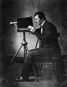
A daguerreotype of gallery owner Jacob Byerly of Frederick, Maryland, operating a typical daguerreotype camera of the early 1840s. From the book “Photography and the American Scene: A Social History, 1839-1889” by Robert Taft, 1938. 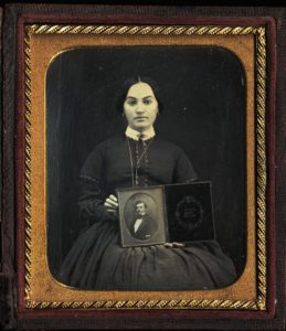
Woman seated, holding full plate daguerreotype portrait of a man, circa 1850. George Eastman House Collection.
Without a doubt daguerreotypes are historical relics, giving us a glimpse into the lives of Victorian people who lived almost 200 years ago. The sensitive nature of this process has caused numerous copper plates to be tarnished and long lost, but μ-XRF offers hope. The technique uses a bright x-ray beam to scan the damaged copper plate. The interaction of the beam with the metals on the surface of the plate produces an element specific signature that allows researchers to map the distribution of these metals with high sensitivity, in this case mercury, uncovering images from the past (see figure). Of interest, μ-XRF microscopy has also revealed “hidden treasures” in Victorian era paintings.
Clearly, scientific methods to recover original images from unrecognizable daguerreotypes are well underway. This innovative approach will surely impact future examinations of copper plate-based antique photograph collections, bringing to light evermore details of the Victorian era.
Peer edited by Jenna Beam